- Наша продукция
- Anesthesia Machines
- Dual automated Gram Stainer
- Biochemistry Analyzers
- Coagulation Analyzers
- Defibrillators
- Dental Units
- Dental X Rays
- Dialysis Machine
- ECG Machines
- Electrolyte Analyzers
- Electrosurgical Units
- ESR Analyzers
- Hematology Analyzers
- Hospital Beds
- Infant Incubators
- Infusion Pumps
- Microplate Readers
- Microplate Washers
- Neonatal Radiant Warmers
- Operating Lights
- Operating Tables
- Patient Monitors
- Phototherapy Lamps
- PPE Kits
- Radiography
- Surgical Suction Pumps
- Ultrasound Scanners
- Urine Analyzers
- Ventilator Machines
- X ray Film processors
- X ray Machines
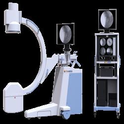
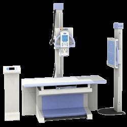
Radiography
Zimed offer C-arm and digital radiographic machines featuring high frequency inverter to emit good quality X-rays at minimal radiation doses. X-ray beam is passed through the body, portion of the x-ray which pass through the internal body structure is transmitted to a detector to record image for later evaluation.
compare product
-
Specification
Fluoroscopic Capacity Tube voltage : 40 to 110 kV
Tube current : 20 to 63 mA
Tube voltage
( Automatic Fluoroscopy ) : 40 kV to 110 kV ( automatic adjustment )
Tube current
( Automatic Fluoroscopy ) : 0.3 to 4 mA
Photographic Capacity Maximum rated capacity : 3.5 kW
Tube voltage and current combination : 40 to 49 kV ; 1 to 125 mAs
50 to 59 kV ; 1 to 110 mAs
60 to 69 kV ; 1 to 90 mAs
70 to 79 kV ; 1 to 80 mAs
80 to 89 kV ; 1 to 71 mAs
90 to 99 kV ; 1 to 63 mAs
100 to 110 kV ; 1 to 40 mAs
mAs : 1 to 125 mAs
X-ray Tube Fixed Anode : Small Focus: 0.3 mm
Large Focus: 1.5 mm
Thermal capacity : 35 kJ
Power supply : 220 V; 50 Hz
Inverter frequency : 40 kHz
Video System Image intensifier : 9 inches
Charged couple camera : Ultra-low light
Monitor : 19 inch LCD
Central control unit : Upright, overturned, positive and negative image
Work station : Yes
Structure Wheel : ± 90 ° rotation
Vertical stand : 400 mm
Orbital rotation : 120 °
-
Features
- Orbital rotation of 120 °
X-ray tube : Small and large focus
Storage of up to 8 high quality images
Optimal bright image resolution with automatic fluoroscopy tracking technique
Minimization of X-ray radiation
Digital workstation for visualization of working parameters
Automatic retention of last captured image
- Orbital rotation of 120 °
compare product
-
Specification
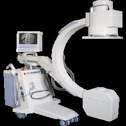
Fluoroscopic Capacity Tube voltage : 40 to 120 kV
Tube current : 20 to 100 mA
Tube voltage
( Automatic Fluoroscopy ) : 40 kV to 120 kV ( automatic adjustment )
Tube current
( Automatic Fluoroscopy ) : 0.3 to 4 mA
Photographic Capacity Maximum rated capacity : 5 kW
Tube voltage and current combination : 40 to 49 kV ; 1 to 180 mAs
50 to 59 kV ; 1 to 140 mAs
60 to 69 kV ; 1 to 125 mAs
70 to 79 kV ; 1 to 110 mAs
80 to 89 kV ; 1 to 100 mAs
90 to 99 kV ; 1 to 80 mAs
100 to 110 kV ; 1 to 63 mAs
110 to 120 kV ; 1 to 50 mAs
mAs : 1 to 180 mAs
X-ray Tube Fixed Anode : Small Focus: 0.3 mm
Large Focus: 1.5 mm
Thermal capacity : 35 kJ
Power supply : 220 V; 50 Hz
Inverter frequency : 40 kHz
Video System Image intensifier : 9 inches
Charged couple camera : Ultra-low light
Monitor : 14 inch CTR attached to C-arm
Central control unit : Upright, overturned, positive and negative image
Work station : No
Structure Wheel : ± 90 ° rotation
Vertical stand : 400 mm
Orbital rotation : 120 °
-
Features
- Orbital rotation of 120 °
X-ray tube : Small and large focus
Storage of up to 8 high quality images
Optimal bright image resolution with automatic fluoroscopy tracking technique
Minimization of X-ray radiation
Digital workstation for visualization of working parameters
Automatic retention of last captured image
- Orbital rotation of 120 °
compare product
-
Specification
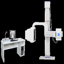
X-ray Machine Power output : 26 kW
X-ray tube (dual focus) : Large focus: 1.3 mm
Small focus: 0.6 mm
Tube voltage : 40 kV to 130 kV
Tube current : 200 mA
Inverter frequency : 60 kHz
Thermal capacity : 900 kJ
Rotatory anode speed : 3000 rpm
mAs : 360 mAs
U-shaped Arm Rotation range : -40 ° to + 130 °
Motorized vertical travel : 450 mm to 1700 mm
Anode to screen range : 1000 mm to 1800 mm
Digital Detector Type : Charge-coupled device ( CCD )
Screen size : 17 × 17 inch
Pixel size : 108 µm
Imaging time : 7 sec
Image storage and transmission : Dicom- 3.0
Radiography bed size : 2000 mm × 650 mm
Photography Bed Bed size : 2000 mm × 650 mm
Bed height : ≤ 720 mm
Transverse shift : 200 mm
Longitudinal shift : 100 mm
Power supply : 380 V ; 50 Hz
-
Features
- Dual focus X-ray tube
Real time microprocessor control
17 by 19 million pixel digital CCD detector
Digital tactile LCD graphic color control system
Radiographic parameters depend upon physical character of patient such as multi-site, multi position, adult and pediatric
Internalized battery allow 200 radiographic exposer on full charge
- Dual focus X-ray tube
compare product
-
Specification
X-ray Machine Tube voltage : 40 kV to 150 kV
Tube current : 10 mA to 630 mA
Focus : 0.6 mm / 1.2 mm
Time : 2 to 6300 ms
Power : 380 V ± 38 V (AC)
Internal resistance : 380 V : 0.5 Ω
Rotating speed : 2800 rpm
Allowed frequency variation : 50 Hz to 60 Hz ± 1 Hz
Digital Detector Screen size : 17 × 17 inch
Image preview time : Not more than 2 sec
Pixel pitch : 127
Special resolution : Not less than 3.9 lp/mm
Radiography Machine Frame Operation mode : Manual / electronic
Horizontal shift : ≥ 250 mm
Vertical shift : ≥ 825 mm
Table tope size : 2100 × 825 × 655 mm
Maximum Load capacity : 250 kgs
-
Features
- Dual focus X-ray tube
Flat panel digital detector
Manual mode operating collimator
High quality grid : 1000 mm to 1800 mm
High voltage cable : 75 kV
- Dual focus X-ray tube
compare product
-
Specification
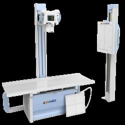
Power output 25 kW Inverter frequency 40 kHz X-ray tube (dual focus) Large focus: ( 1.3 × 1.3 ) mm
Small focus : (0.6 × 0.6 ) mmTube current 200 mA Rotatory anode speed 3000 rpm Tube voltage 40 kV to 125 kV Power supply 200 mA X-ray tube rotation 380 V; 50 Hz Table size ± 90 ° Table height 2000 mm × 760 mm Transverse travel of table ≤ 700 mm Longitudinal travel of table ±110 mm (Electromagnetic brake) Floor stand rotation ±325 mm (Electromagnetic brake) Floor stand longitudinal travel 360 ° Axial rotation ≥ 1350 mm SID (Source to image distance) 0 to 35 ° -
Features
- High frequency and high voltage generator
Radiographic parameters depend upon physical character of patient such as multi-site, adult, pediatric
Bucky stand for head, chest, belly, pelvic cavity and backbone imaging
Radiographic table with electromagnetic brake is movable, guarantees the accurate and simple position
LCD touch screen graphic control alters radiographic parameters
Automatic fault detection and safeguard system
- High frequency and high voltage generator